Definition: MALT refers to mucosal associated lymphoid tissues. The MALT lymphoma is a proliferation of monocytoid B cells. The concept is that this type of lymphoid proliferation arises from lymphoid tissue that is normally found in mucosa such as the conjunctiva and GI tract.
Etiology: Recent studies suggest Chlamydia psittaci is responsible for conjunctival MALT lymphoma, and showed regression of ocular adnexal lymphoma after antibiotic therapy. The organism is found in about 80% of cases by PCR (
Reference 1).
Incidence/Prevalence: By far this is the most common lymphomas of the conjunctiva and orbit. Previously considered as atypical lymphoid proliferations that could progress, the designation as MALT lymphoma permits the merging of MALT with the mainstream lymphoma classifications. There is a clear female predominance and older population for MALT.
Clinical Findings: These tumors may arise in the conjunctiva, eyelid, lacrimal gland and orbit. Generally, occurring in older individuals, more common in women, MALT lymphoma manifests in the orbit as a mass that molds around the eye or the so-called fish flesh or salmon patch under the conjunctiva. MALT lymphomas are generally insidious, indolent, progress slowly, and are characterized by recurrence many years following the initial diagnosis.
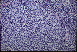
Histopathology: MALT Lymphoma is characterized by poorly defined follicular appearing areas that are composed of monocytoid B cells that feature enlarged nuclei. Click on the image below to enlarge.
 At the periphery, lymphocytes appear smaller and sport round regular nuclei. Often the cytoplasm has a water clear appearance. The infiltrates are heterogenous with T cells, small lymphocytes, plasma cells and a variety of reactive lymphoid cells. Critical to the correct diagnosis is the establishment of a proliferation of atypical monocytoid B cells that expand and infiltrate the normal tissue. Immunohistochemistry marks the proliferating cells as B cells, CD20 positive and negative for CD 10 and CD 5. The cells may be CD 43 (sialophorin) positive and may show light chain restriction. Gene rearrangement studies will often show clonal gene rearrangement of B cell gene.
Treatment: Low dose radiotherapy in multiple courses with proper shielding has been the traditional treatment but recently some groups have reported success with chlorambucil. Other groups have noticed regression with doxycycline 100 mg bid x 3 weeks.
Prognosis: The prognosis is excellent with a 10 year survival of 73-86% and very few if any patients dying of MALT. However, many patients recur and there can be transformation to a more malignant lymphoma such as a large B cell lymphoma. Spontaneous regression of tumor may obfuscate treatment assessment.
References:
1. Ferreri et al. J Clin Oncology 2006; 23:5067
2. Ben Simon et al. Opthalmology 2006; 113:1209.
3. Suh et al. Int J Radiat Oncol Biol Phys. 2006 65;228-33. Orbital marginal zone B-cell lymphoma of MALT: radiotherapy results and clinical behavior.
